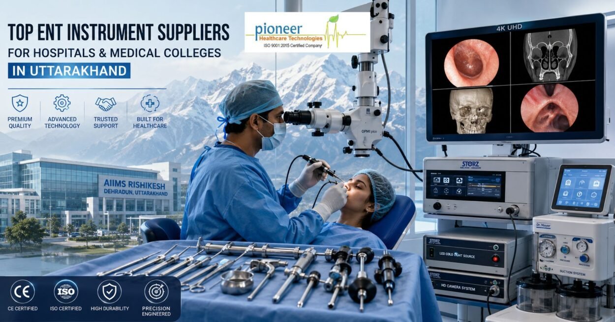
for decades, the standard joke among operating room staff was that surgeons needed a hybrid of X-ray vision and supreme guesswork to navigate the narrow pathways of the human body. Early iterations of rigid endoscopy—while revolutionary for their time—often forced clinicians to peer through a grainy, two-dimensional lens, relying heavily on tactile feedback and shadow cues to distinguish between a critical nerve and a minor blood vessel.
The baseline surgical paradigm has fundamentally changed. Driven by rapid consumer electronic breakthroughs and hyper-advanced optical engineering, the operating room is experiencing a visual renaissance. The simultaneous integration of Ultra-HD 4K resolution and 3D stereoscopic visualization into rigid endoscopic systems is transforming how minimally invasive surgeries (MIS) are performed.
From ENT and neurosurgery to complex laparoscopy, these advanced display technologies are moving beyond luxury upgrade status to become the absolute clinical standard for patient safety and procedural efficiency.
The Monocular Trap: Moving Past Traditional 2D Imaging
To understand why 4K and 3D are winning the market, we must first address the limitations of traditional Full HD (1080p) 2D imaging.
When a surgeon views a three-dimensional internal organ on a flat, two-dimensional monitor, they lose binocular depth perception. To compensate, they have to rely on indirect, monocular depth cues—such as instrument shadows, the relative size of anatomical structures, and perspective movement.
While experienced, board-certified surgeons adapt well to this “monocular trap,” it drastically steepens the learning curve for residents and introduces avoidable cognitive fatigue during grueling, hours-long procedures. Furthermore, in tight, structurally complex fields like the nasal passages or the skull base, a lack of precise spatial depth can dramatically increase the risk of accidental tissue perforation or incomplete lesion resections.
The 4K Advantage: The Power of Spatial Resolution
Ultra-HD 4K technology delivers a native resolution of 3840 × 2160 pixels, a massive fourfold increase over traditional standard Full HD predecessors. This dramatic boost in pixel density fundamentally reshapes the surgical environment in several tangible ways:
- Micro-Vascular Clarity: High-performance 4K systems leverage a significantly wider color gamut. This allows surgeons to perceive ultra-fine gradations in reds and pinks, making it infinitely easier to distinguish between distinct mucosal boundaries, delicate nerve pathways, and minute capillary networks.
- Digital Zoom Efficiency without Pixelation: Because 4K captures four times the data of a standard screen, surgeons can digitally zoom in on a targeted lesion or bleeding site without experiencing blurry, pixelated artifacts. This allows the camera head to remain at a safer distance from the active surgical site, reducing the frequency of lens fouling from blood splatter or condensation smoke.
- Indirect Depth Cues via Shading: Clinical studies evaluating narrow-space surgical maneuvers demonstrate that 4K’s enhanced color contrast and high-dynamic-range (HDR) processing engine provide exceptional indirect depth cues. By highlighting microscopic shadows and surface textures, 4K mitigates a significant portion of the spatial loss inherent to traditional flat screens.
The 3D Paradigm: Restoring Human Depth Perception
While 4K maximizes resolution, 3D stereoscopic vision restores the natural, binocular depth perception that open surgery famously provides. Utilizing modern dual-lens rigid endoscopes paired with passive circular polarizing glasses, 3D systems transmit distinct images to the surgeon’s left and right eyes simultaneously.
The immediate clinical impact is profound. Large-scale surgical trials, including the notable IDOSP randomized cross-over study, have rigorously compared 3D display systems against 4K setups in minimally invasive environments. The findings underscore an undeniable trend: 3D visualization optimizes surgical performance across the board.
| Parameter | Impact of 3D Over 2D-4K | Clinical Realization |
|---|---|---|
| Execution Time | Shorter performance times across standard surgical tasks (e.g., peg transfer, precision suturing). | Reduced overall anesthesia time for patients. |
| Error Rates | Noticeably lower mistake frequencies, particularly during complex, depth-dependent movements like intracorporeal knot tying. | Enhanced patient safety and lower complication rates. |
| Cognitive Load | Measured via the NASA-TLX index, mental fatigue dropped significantly for operating surgeons. | Reduced surgeon exhaustion during high-stakes, long-duration procedures. |
Interestingly, the data reveals that while seasoned, board-certified surgeons utilize their deep spatial memory to navigate effectively on 4K systems, medical students and non-board-certified residents see the highest performance jumps when switching to 3D. It effectively flattens the steep learning curve of minimally invasive techniques, allowing younger clinicians to master complex orientation tasks with significantly greater confidence.
4K 3D Hybrid Systems: The Convergence Era
For a brief period, hospital procurement departments faced a difficult dilemma: do we choose the unparalleled spatial clarity of 4K, or the anatomical depth perception of 3D?
Today, the industry has bypassed that compromise. Leading medical manufacturing giants have launched fully integrated 4K 3D rigid endoscopic camera towers. These state-of-the-art platforms utilize dual ultra-high-definition CMOS sensors embedded directly into an ergonomic, lightweight camera head.
By combining an expansive depth of field with cinematic 4K detail and true stereoscopic depth, these systems create an immersive visual ecosystem. When integrated with advanced near-infrared (NIR) and indocyanine green (ICG) fluorescence imaging modalities, a surgeon can visualize real-time blood perfusion and lymphatic structures overlaid perfectly onto a high-definition 3D map of the patient’s anatomy.
The Modern Ecosystem: AI-Driven Surgery
The rise of 4K and 3D imaging serves another crucial purpose: it lays the necessary groundwork for digital surgery ecosystems and machine learning.
Artificial intelligence algorithms thrive on high-fidelity, data-dense inputs. The crystal-clear feeds generated by modern 4K camera control units provide the perfect raw material for real-time edge computing. Today’s cutting-edge towers are beginning to implement onboard deep learning models capable of:
- Dynamically flagging suspicious tissue anomalies and early-stage margins.
- Providing real-time anatomical tracking and spatial boundaries during delicate sinus or skull-base dissections.
- Automatically adjusting lighting and stabilizing camera shake during high-stakes maneuvers.
Conclusion
The evolution of rigid endoscopy from blurry monitors to crystal-clear 4K 3D ecosystems represents a monumental victory for patient outcomes. By bridging the gap between open-surgery depth perception and minimally invasive precision, these systems empower surgical teams to work faster, make fewer errors, and navigate the human body with unmatched confidence.
For hospital administration and medical procurement officers looking to future-proof their operating theaters, the decision is clear: investing in premium 4K and 3D visualization is no longer an optional luxury—it is the modern benchmark for patient care.
Author:- ENT instrument by pioneermedical
Leave a Comment